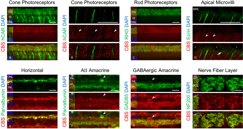
Figure 8.
Immunohistochemical analysis of canine retinal cryosections labeled with CBS and retinal cells markers. Retinal cryosections immunolabeled with anti-CBS (red), DAPI (blue), hCAR (green), rhodopsin (green), ezrin ( green), parvalbumin (green), GAD65 (green) and NFH (green). Results showed that CBS was not localized in cones (A) and confocal microscopy analysis of section immunolabeled with hCAR and CBS confirmed that cones did not express CBS (arrow heads shows the locations of cones) (B). Double immunnolabeling with rhodopsin (a marker for rods outer segment) and CBS confirmed localization of CBS in rods outer segment (C). Confocal analysis of section showed that RPE apical microvilli illustrated by ezrin labeling show lack of CBS in cones (arrow heads) but highlight CBS expression associated with the rods (arrows) (asterisks show the nucleus for RPE) (D). The results also confirmed localization of CBS in horizontal (E), AII amacrine (arrow head shows the cell bodies and dendrites are labeled by asterisk) (F) and GABAergic amacrine cells (arrow heads shows the cell body and dendrites are labeled by asterisk) (G) and NFL (H). Because the parvalbumin labeling (green) in amacrine’s dendrites is weaker than CBS (red) in AII amacrine cells, the green signal has been enhanced to illustrate colocalization (Scale bar: 20 µm).

