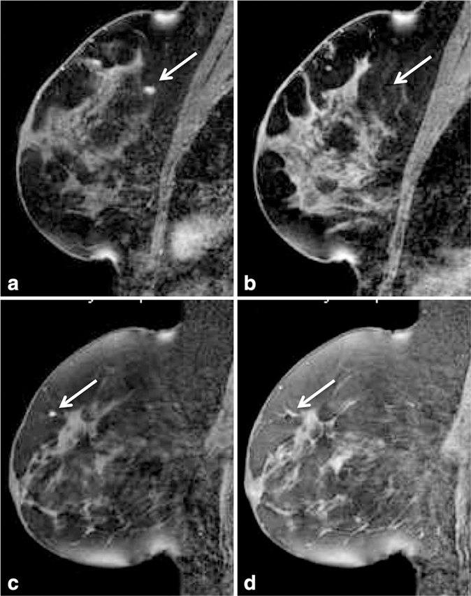
Figure 16.
Alteration of fat-suppression to improve visualization of obturator needle tip during MR guided biopsy. Sagittal T1-weighted images with fat suppression after administration of IV gadolinium are shown for two patients. In the first patient (top row), lesion identification scan (a) demonstrates excellent visualization of a 5 mm enhancing mass (arrow) at posterior depth in the superior breast surrounded by adipose tissue. Scan to confirm needle location before biopsy (b) demonstrates the challenge in identifying the obturator tip (arrow) with fat-suppression settings unaltered. In this case, the tip was identified to be within adipose tissue 5 mm superficial to the enhancing mass. In the second patient (bottom row), lesion identification scan demonstrates excellent visualization of a 3 mm enhancing focus (c) at anterior depth in the superior breast surrounded by adipose tissue. By decreasing the level of fat suppression for the needle-confirmation images (d) through alteration of the SPAIR delay, improved visualization of the obturator tip (arrow) was achieved and confirmed to be within the enhancing focus.

