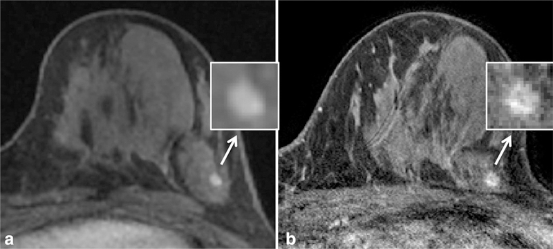
Figure 3.
Example of improved spatial resolution from dynamic contrast enhanced MRI at 1.5T (a) to 3T (b) leading to a change in lesion classification in a high-risk patient. At 1.5T, the lesion was described as a 4 mm oval circumscribed mass (arrow) with smooth margins (inset), probably benign (BI-RADS category 3). At 3T, the margins demonstrated fine spiculations (inset), and the mass was re-categorized as suspicious (BI-RADS category 4). MR guided biopsy yielded invasive ductal carcinoma. Detailed scan parameters for breast MRI at 1.5T and 3T at the University of Washington are summarized in Table 3.

