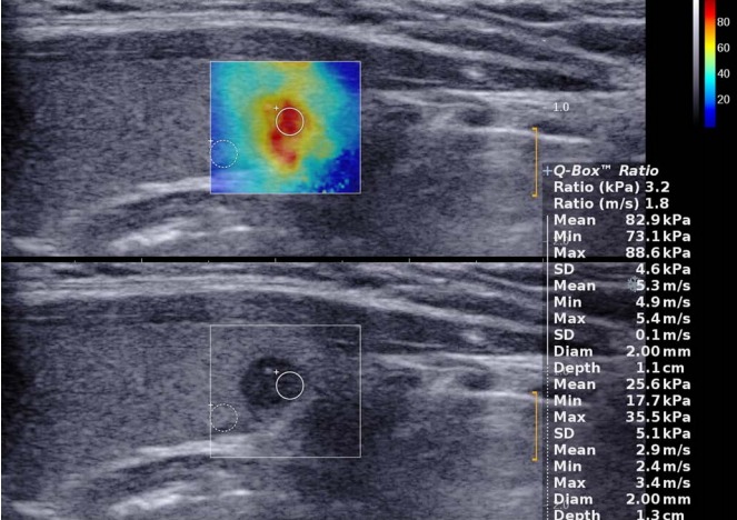
Fig. 12. Papillary thyroid carcinoma in a 28-year-old woman.

Gray-scale ultrasonography (lower side) shows a solid hypoechoic 6-mm thyroid nodule with poor margin and microcalcifications. SuperSonic shear-wave elastography (upper side) shows a heterogeneously stiff (red and yellow) nodule with a maximum elasticity of 88.6 kPa.
