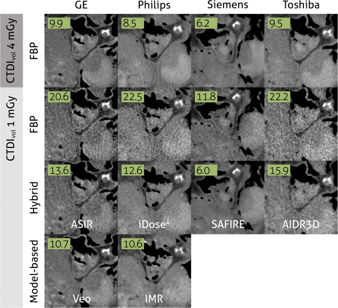
Fig. 3.
One ex vivo human heart, scanned at 4 mGy and 1 mGy (75% dose-reduction) with high-end CT scanners from four vendors. Images are reconstructed with filtered back projection (FBP), hybrid iterative reconstruction, and model-based iterative reconstruction. Numbers represent noise levels (standard deviations) in air. Images derived from a study published before by Willemink et al [39]

