FIGURE 3.
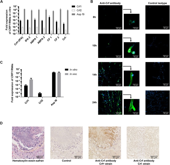
Expression of Crf transcripts and isoforms in clinical strains, in vitro and in vivo. (A) CRF1 RNA transcript expression in different clinical strains, by RT-qPCR. Results were normalized toward gpdA and TUB1 housekeeping genes, set to 1. Results are expressed in mean ± SD. IPA, strains from patients with invasive pulmonary aspergillosis (including Crf+); ABPA, strains from patients with allergic bronchopulmonary aspergillosis; CF, strains from patients with colonization (cystic fibrosis); Crf-, clinical strain with natural frameshift mutation, with no expression of Crf proteins. (B) Expression kinetic and localization of Crf proteins (in green) in vitro by immunofluorescence in confocal microscopy. MS112-IIB1 scFv-Fc anti-Crf antibody (or isotype) at 2 μg/mL were used. Cell nuclei were stained in Hoechst (in blue). Scale bar: 50 μm or 10 μm. Original magnification: ×400. (C) Differential expression of CRF1 RNA transcript in vivo and in vitro, by RT-qPCR. RNAs were extracted Crf+ strain cultured in vitro or from lungs of rats infected by Crf+ strain. Results (n = 5) are expressed in mean ± SD. (D) Expression of Crf proteins (in brown) in vivo, in rat lungs tissues, by immunohistochemistry. Slides of rat lungs either infected by Crf+ or Crf- (natural mutant) strain were studied with MS112-IIB1 scFv-Fc antibody at 2 μg/mL or control. One slide was treated in HES coloration. Scale bar: 50 μm. Magnification: ×400.

