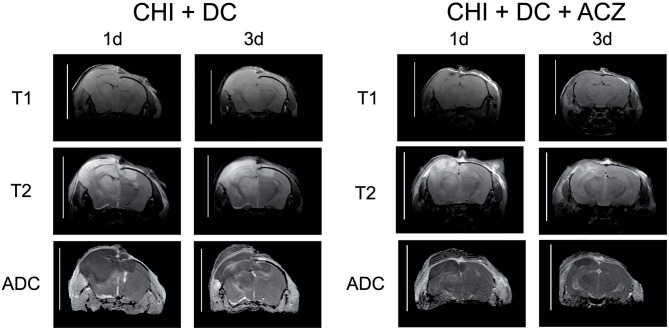
Figure 5.
Illustrating the difference in brain edema course, observed between the animals treated with acetazolamide after trauma/decompression and between placebo-treated group. MRI-scans (T1 sequence, T2 sequence, and apparent diffusion coefficient (ADC) map) were obtained from animals subjected to closed head injury followed by decompressive craniectomy (CHI+DC) without acetazolamide (ACZ) administration and from animals with additional i.p. administration of ACZ (CHI+DC+ACZ). The scanning was performed 1d and 3d postinjury. The animals with craniectomy applied after CHI presented significant amount of brain edema. Already 1d postinjury, a part of edema displayed vasogenic properties, as expressed by hyperintensity in ADC map sequence. In contrast, in animals with additional ACZ treatment both cytotoxic (hypointense) and, strikingly, vasogenic (hyperintense) edema was less manifest at both analyzed time points. The scans were obtained in representative animals using 9.4 T MRI scanner; bar = 10 mm.

