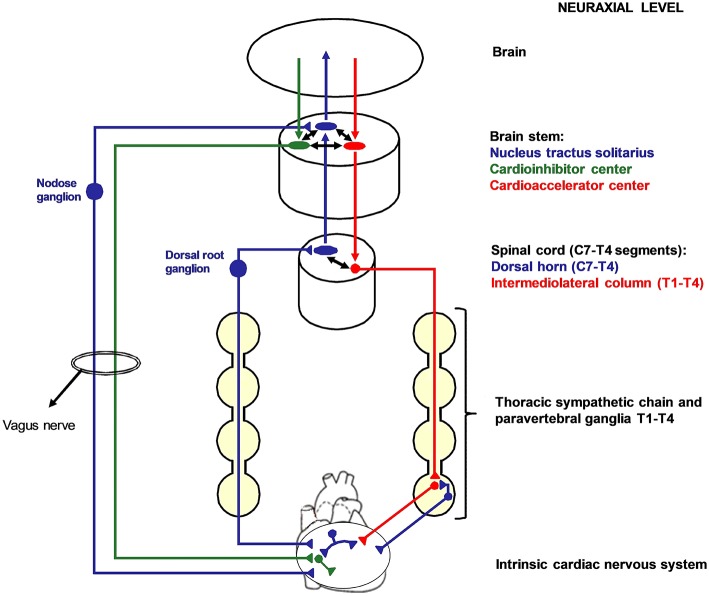
Figure 1.
Cardiac nervous system organization in humans. Blue: afferent nervous system with its ganglia: nodose ganglia and C7-T4 dorsal root ganglia (DRG). Green: parasympathetic efferent nervous system. Red: sympathetic efferent nervous system. All the afferent and efferent structures outside the central nervous systems are bilateral, although mostly represented as unilateral for simplicity. Cardiac afferent fibers traveling across the paravertebral sympathetic ganglia (usually referred to as cardiac sympathetic afferent fibers) directly reach the DRG without having synapsis before. These fibers mediate cardio-cardiac sympathoexcitatory spinal reflexes that significantly increase the sympathetic output to the heart. Left cardiac sympathetic denervation (LCSD) consists in the removal of the left thoracic sympathetic chain and paravertebral ganglia from T1 to T4. Since ipsilateral DRG are spared by LCSD, a left afferent reinnervation from the DRG to the heart is theoretically possible with time. On the other hand, the left efferent sympathetic system from T1 to T4 is interrupted at a preganglionic level; therefore, no ipsilateral efferent sympathetic reinnervation is possible after LCSD.

