Figure 4.
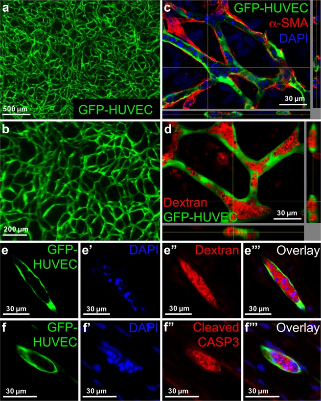
Substitution of Matrigel/rCOL by hCOL from fibroblasts leads to lumenized EC network formation with involvement of apoptosis. (a,b) 3D hydrogel constructs containing GFP positive HUVECs and hASCs and hCOL after 14 days of cultivation in SFM. (c) Virtual stacks of GFP-HUVECs co-cultured with hASCs in hCOL for 14 days and stained for α-SMA. Nuclei were counterstained with DAPI. α-SMA positive cells are in physical contact with GFP-HUVECs. (d) Virtual stacks of HUVECs co-cultured with hASCs and incubated with Texas red-labeled Dextran. (e–e”’) Images of cryo-sections from constructs containing GFP-HUVECs, hASCs and hCOL. Constructs were cultivated for 14 days in SFM and incubated with Texas red-labeled dextran. Cryo-sections were stained with DAPI. (e) GFP-HUVEC, (e’) DAPI, (e”) Texas red-labeled dextran, (e”’) Superimposed image of (e,e’,e”). Intense DAPI staining of fragmented DNA is localized within the lumen filled with Texas red-labeled dextran. (f–f”’) Images of cryo-sections from constructs containing GFP-HUVECs, hASCs and hCOL. Constructs were cultivated for 14 days in SFM. Cryo-sections were stained for cleaved caspase 3 and DAPI. (f) GFP-HUVEC, (f’) DAPI, (f”) cleaved caspase 3 (CASP3), (f”’) superimposed image of (f,f’,f”). Intense DAPI staining of fragmented DNA is localized within the lumen and co-localizes with the signal for cleaved caspase 3. Scale bar: (a) 500 µm; (b) 200 µm; (c–f”’) 30 µm.

