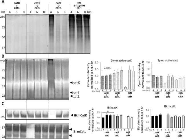
Figure 5.
Soluble tendon ECM fragments stabilize catK and catL while being degraded, and catK is primarily responsible for tendon ECM degradation. Concurrent and sequential catK and catL were incubated with soluble tendon ECM for 4, 6, and 8 hrs. SDS-PAGE showed the soluble tendon ECM remaining after incubation time course. At all time points with either concurrent co-incubation or when primed with catK before the addition of catL, ECM fragments were below Coomassie detection. Soluble tendon ECM primed with catL was degraded only after catK was added, with fragments still visible even by 8 hrs. (A) Soluble tendon ECM sustained active catK and catL by zymography at all three time points. CatK was active and associated with tendon ECM fragments at multiple molecular sizes throughout the zymography gel. (B) Densitometry is shown on the right, and there is no significant difference in signals for these time points except for 4 and 8 hours K + L condition, (n = 3, p < 0.05). Similarly, by Western blot catK and catL were detected at similar levels at all three time points where added, with densitometry shown on the right. (C) Representative gels and images shown from three biological replicates.

