
Figure 3.
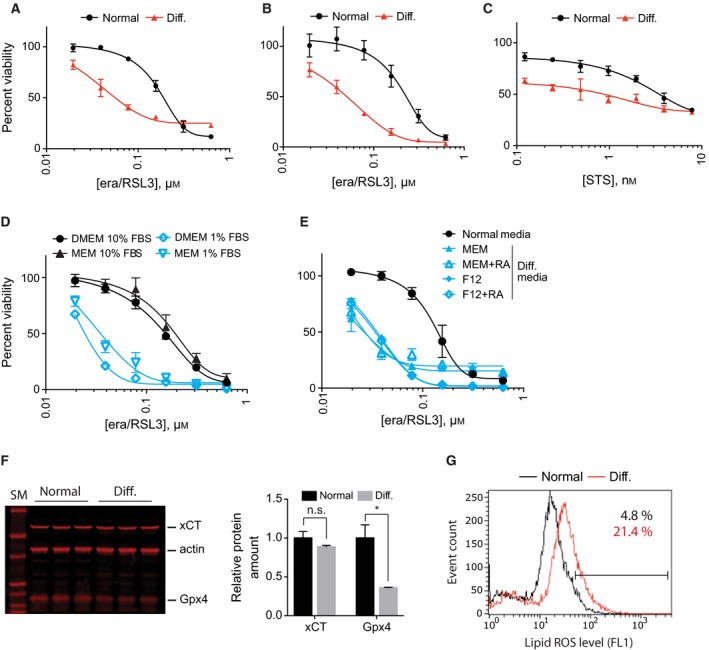
NSC‐34 cells became more sensitive to ferroptosis upon differentiation. (A) Comparison of ferroptosis sensitivity between NSC‐34 cells grown in normal and differentiation media. Cell viability was determined by fluorimetry using resazurin dye. (B) Same as in (A) except that cell viability was determined by luminometry using RealTime‐Glo reagent. (C) Upon differentiation, NSC‐34 cells became sensitive to staurosporine treatment as well, which indicates that sensitization effect is not specific to ferroptotic cell death. (D) Low serum (1%) in differentiation media caused ferroptosis sensitization in NSC‐34 cells. (E) NSC‐34 cells became sensitive to ferroptosis in all four differentiation conditions because they all contain low serum. NSC‐34 cells were treated with indicated amount of lethal compounds for 24 h. Cell viability in (A–E) was determined using resazurin viability dye. Data are presented as mean ± SD; n = 3. (F) Western blot analysis showed GPx4 expression was decreased in differentiation condition while the xCT level did not change. Quantifications of the results are shown in the graph. Data are presented as mean ± SD; n = 3. n.s., not significant; *P < 0.05 by Welch's t test. (G) NSC‐34 cells showed higher level of lipid peroxides in differentiation condition. BODIPY‐C11 dye was used to determine lipid peroxide level.