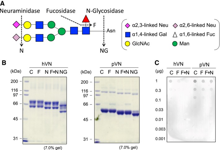
Figure 8.

Preparation of hVN and pVN treated with glycosidases. (A) Cleavage site of enzymatic deglycosylation in the structure of the fucosylated N‐glycan of hVN and pVN. (B) Glycosidase‐treated hVNs and pVNs (4 μg per lane) were loaded in each lane of 7.0% polyacrylamide gel and run for SDS/PAGE under reducing conditions with 2‐mercaptoethanol. The gels were stained with CBB. (C) Dot blot analysis of PVDF membrane stained with the biotinylated UEA‐I for detecting Fuc in VNs. C, control VN incubated without enzyme; F, VN treated with fucosidase; N, VN treated with neuraminidase; F+N, VN treated with both fucosidase and neuraminidase; NG, VN treated with N‐glycosidaseF.
