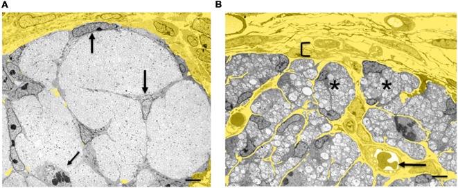
Figure 4.
Architectural reorganization of developing nerves. (A) Electron micrograph of a transverse section through E14 rat sciatic nerve. SCP are embedded among the axons and at the surface of the nerve (big arrows). A dividing SCP is also seen (small tilted arrow). Note that connective tissue (yellow) surrounds the nerve, but is not found inside the nerve. (B) Electron micrograph of a transverse section through E18 rat sciatic nerve. iSch surround the collection of axons, forming compact groups (“families”; examples indicated by asterisks). Extensive connective tissue (yellow) containing blood vessels (arrow) is now found inside the nerve surrounding the families, as well as outside the nerve. Bracket indicates the developing perineurium (scale bar: 4 μm).

