Figure 6.
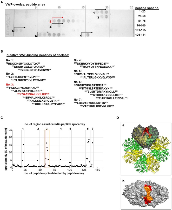
Identification of a putative VWF binding site on the pneumococcal enolase. (A) VWF overlay of enolase peptide spot array, representing the whole enolase amino acid sequence, identified up to seven regions displaying VWF-binding activity. Antibody controls are shown in Figure S1B. (B) Table of peptide sequences of all peptide spots showing VWF-binding activity. The strongest VWF-binding signal shows peptide “181YGAEIFHALKKILKS195” of region no. 3 (marked in red). (C) Densitometric quantification of signal intensity. The seven regions with positive signals are numbered and marked with squares. Peptide region Nr. 3 is located on the enolase molecule surface and the core peptide is marked in red and the adjacent peptide spots are marked in orange. (D) Localization of the putative VWF-binding pocket within the octameric enolase molecule depicts alternating enolase monomers colored in yellow and green; the VWF-binding site is highlighted within one of the four dimers colored in gray (a). The whole VWF-binding peptide region no. 3 representing aa 181–195 is marked in orange and the core peptide composed of aa 181–195 is marked in red. Top view of the VWF binding groove of a dimer is shown in (b). Additional views are added in Figure S5. Structure visualization was calculated using PyMOL.

