. 2019 Mar 25;49(1):53–58. doi: 10.5624/isd.2019.49.1.53

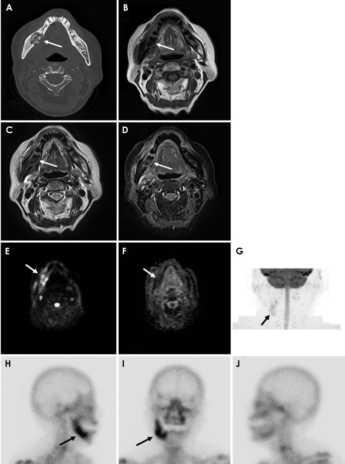
Fig. 1. Medication-related osteonecrosis of the right side of the mandible in an 86-year-old woman. A. Axial bone tissue algorithm computed tomography shows an osteolytic lesion with sequestrum separation and periosteal bone proliferation in the right mandible (arrow). B. On magnetic resonance imaging, an axial T1-weighted image shows heterogeneous, low-signal intensity (arrow). C. An axial T2-weighted image shows heterogeneous, low-signal intensity (arrow). D. An axial short TI inversion recovery image shows heterogeneous, high-signal intensity (arrow). E. An axial diffusion-weighted image shows heterogeneous, high-signal intensity (arrow). F. An axial apparent diffusion coefficient map shows heterogeneous, low-signal intensity (arrow). G. A maximum intensity projection (diffusion-weighted image) shows the lesion in an improved way (arrow). H–J. Bone scintigraphy shows increased uptake in the mandible (arrow).

Fig. 1