Figure 3.
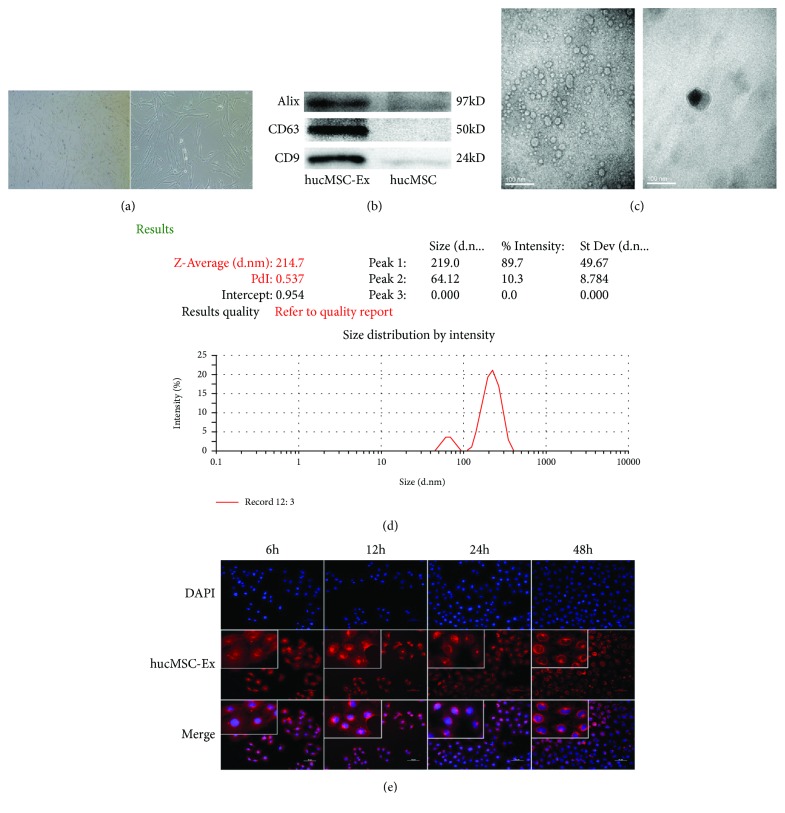
Characterization and internalization of hUC-MSC exosomes. (a) Morphology of hUC-MSCs in exo-free medium. (b) Western blotting for exosome markers in hUC-MSC exosomes and hUC-MSCs. (c) Morphology of hUC-MSC exosomes under transmission electron microscopy. (d) NTA of hUC-MSC exosomes. (e) Fluorescence microscopy of HK-2 cells and hUC-MSC exosomes after coincubation for 6, 12, 24, and 48 h (left to right). These results were representative of at least three independent experiments.

