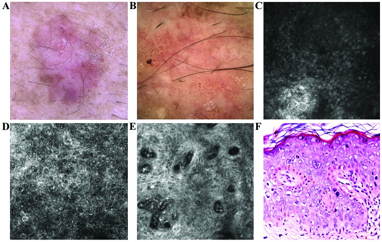
Figure 2.
(A) Clinical image of a flat plaque showed pink and brown diffuse areas. (B) Corresponding dermoscopic image showed diffuse, structureless brown pigmentation, some brown globules and an atypical vascular pattern consist of glomerular, dotted and linear vessels. (C) RCM image (500×500 µm) at the level of the stratum corneum where parakeratotic cells were identified as round to polygonal, refractile structures. (D) RCM image (500×500 µm) revealed the atypical honeycomb pattern in the spinous layer, with various shapes and sizes of the cells and nuclei. Many of these cells have a target-like aspect, with a bright center and a dark peripheral halo. Another type of cell with targetoid aspect, with a dark center and a bright peripheral rim, was also identified in smaller numbers. (E) RCM image (500×500 µm) at the level of the dermo-epidermal junction showed enlarged dermal papillae, with bizarre shapes, without the papillary rings of basal cells, inside of which are identified dilated capillaries, some captured in horizontal section, parallel to the skin. (F) Histopathological image displaying important cytonuclear pleomorphism with hypertrophy and nuclear monstrosities and atypical mitosis at different levels in epidermal thickness (hematoxylin and eosin staining; ×200 magnification).

