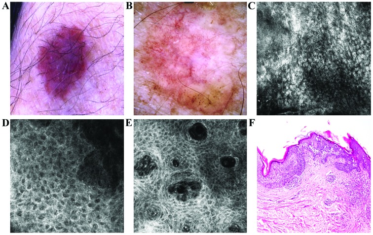
Figure 3.
(A) Clinical image of a slightly variegated brown plaque with very fine scaling. (B) Dermoscopic view showed structureless asymmetric brown pigmentation with erythematous background, few brown globules and irregular network in the periphery and an atypical vascular pattern. (C) RCM examination (500×500 µm) revealed parakeratosis in the horny layer. (D) RCM image (500×500 µm) at the level of the spinous-granular layers showed a disarrayed pattern and target-like cells. (E) RCM image (500×500 µm) at the dermo-epidermal junction the dermal papillae are filled with many dilated capillaries. (F) Corresponding histology with epidermal thickening due to proliferation of atypical squamous cells with cyto-nuclear atypia extended through the whole thickness of the epidermis (hematoxylin and eosin staining; ×100 magnification).

