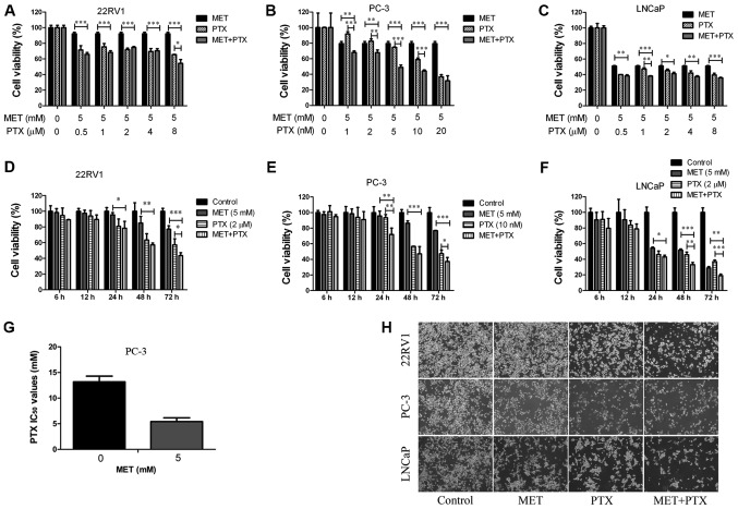
Figure 2.
MET in combination with PTX suppresses cell proliferation. (A-C) Prostate cancer cells were treated with MET (5 mM) and PTX (1, 2, 5, 10, 20 nM for PC-3 cells, and 0.5, 1, 2, 4, 8 µM for 22RV1 and LNCaP cells) for 48 h, and viability was measured by MTT assay. (D-F) Cell viability was measured by MTT assay following treatment with MET (5 mM) and PTX (10 nM for PC-3 cells, and 2 µM for 22RV1 and LNCaP cells) for 6, 12, 24, 48 and 72 h. (G) Changes in IC50 of PTX following MET treatment in PC-3 cells. (H) 22RV1, PC-3 and LNCaP cells were treated with MET (5 mM), PTX (10 nM for PC-3 cells, and 2 µM for 22RV1 and LNCaP cells) and MET + PTX for 24 h. Images of cells were captured using inverted contrast microscopy (magnification, ×100). Cells treated with DMSO were used as the control group with cell viability set at 100%. *P<0.05, **P<0.01, ***P<0.001. DMSO, dimethyl sulfoxide; IC50, half maximal inhibitory concentration; MET, metformin; PTX, paclitaxel.

