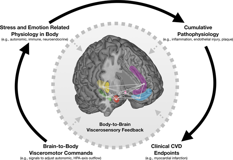
Figure 1.

Neuroanatomically connected limbic and cortical brain regions linking negative emotion, psychological stress, and regulation of peripheral physiology. Highlighted are limbic regions including the amygdala (red), hippocampus (green), and hypothalamus (pink) as well as cortical regions including the insula (yellow), anterior cingulate cortex (purple), and ventromedial prefrontal cortex (blue). Negative emotion and psychological stress engage these regions via changes in (1) local (within-region) activity, (2) distributed and patterned (across-region) activity, and (3) network-level interactions between regions across over time. These responses issue brain-to-body visceromotor commands via specific brainstem nuclei to influence physiology in peripheral organs. Exaggerated or prolonged engagement of these responses promote cumulative pathophysiology and future clinical CVD endpoints. Along this pathway, the effect of stress and negative emotion on the body affects the brain via body-to-brain viscerosensory feedback (dotted arrows), including baroreceptor firing, recruitment of circulating mediators of systemic inflammation into the brain, and brain structural damage/remodeling following myocardial infarction.
