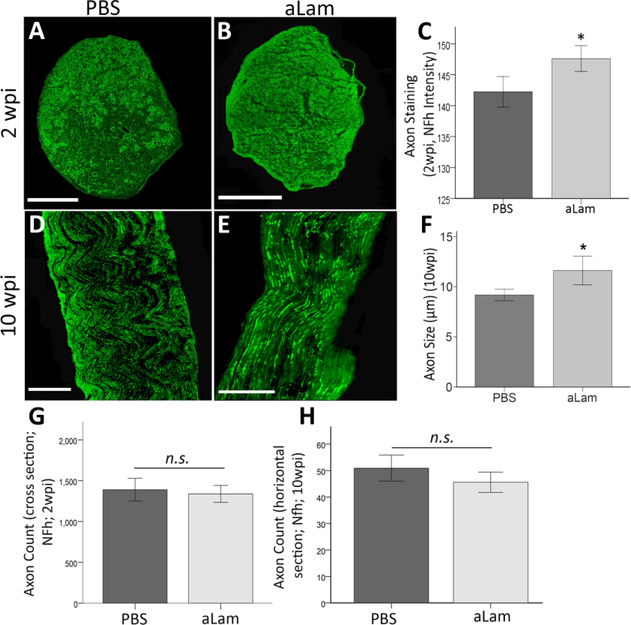
Fig. 5.
Axonal response to aLam. aLam treatment increased axon presence and maturation after injury. A/B) Cross section of injured nerve at 2 wpi treated with PBS (A) or aLam (B) and stained with antibodies recognizing NF-h (α-RT97, green); C) Bar graph illustrating significantly higher axon (NF-h) fluorescent staining intensity (integrated density) on tissue cross sections from the aLam treated group at 2 wpi (Student T-test, ⋆p = 0.04); D/E) Longitudinal section of injured nerve at 10 wpi treated with PBS (D) or aLam (E) and stained with antibodies recognizing NF-h (α-RT97, green); F) Bar graph illustrating significantly increased axon diameter (lm; NF-h particle diameter) with aLam treatment at 10 wpi (Student t-test, *p = 0.01). G/H Bar graphs illustrating similar numbers (counts) of NF-h+ axon at 2 and 10 wpi. Error bars represent SEM. (For interpretation of the references to colour in this figure legend, the reader is referred to the web version of this article.)

