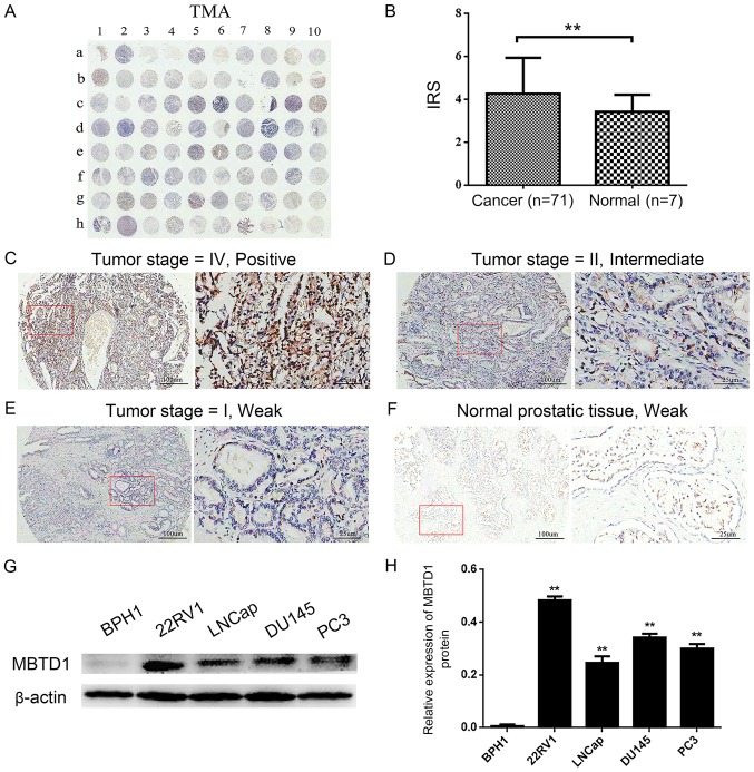
Figure 1.
Overexpression of MBTD1 in PCa tissues and cell lines. (A) IHC staining of MBTD1 in the tissue microarray cohort. (B) The differences in immunoreactivity scores of MBTD1 between PCa tissues and normal prostate tissues. **P<0.01. IHC staining of the distribution of MBTD1 in the cytoplasm of PCa and the different intensities of MBTD1 designated (C) positive, (D) intermediate and (E) weak. (F) Weak staining of MBTD1 in normal prostate tissues. The magnification in the left and right panels is ×40 and ×200, respectively. Representative (G) western blots and (H) quantification of the expression of MBTD1 protein in 22RV1, LNCap, DU-145, PC3 and BPH1 cell lines. **P<0.01 vs. BPH1. PCa, prostate cancer; MBTD1, malignant brain tumor domain containing protein 1; IHC, immunohistochemistry.

