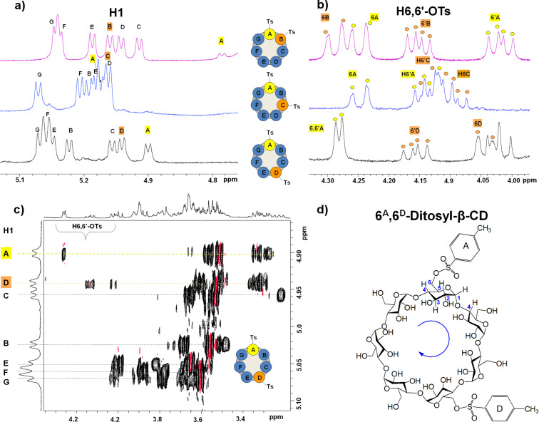
Figure 3.
NMR spectral regions of the three ditosyl regioisomers in D2O (500 MHz). The signals of the tosylated glucopyranose units are indicated in yellow and orange in each spectrum: 1H NMR spectrum of all ditosyl derivatives indicating (a) the anomeric proton (H1) region (5.20 to 4.70 ppm) and (b) the H6,6’-OTs proton region (4.30 to 3.90 ppm). (c) 2D TOCSY NMR spectrum of 6A,6D-ditosyl-β-CD: starting from each H1 resonance, identification of the signals that belong to the same spin system (dotted lines) is possible leading to recognition and assignment of each of the glucopyranose units. (d) Scheme for the clockwise connectivity in 6A,6D-ditosyl-β-CD.

