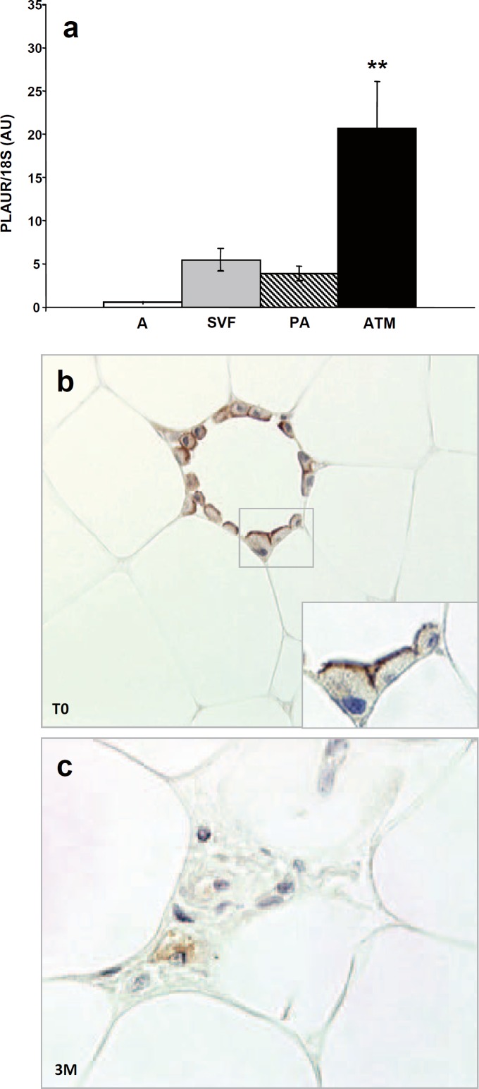
Fig. 2.
a PLAUR gene expression (by RTqPCR) in mature adipocytes (A), in the adipose tissue stroma vascular fraction cells (SVF), in isolated preadipocytes (PA) and in isolated adipose tissue macrophages (ATM) of obese subjects (n = 5). PLAUR expression values were normalized on 18S expression levels and indicated as arbitrary units (AU); ** p value < 0.01 ATM versus A, SVF and PA. b Detection of PLAUR protein in subcutaneous WAT by immunohistochemistry. PLAUR immunopositivity concentrated on adipose tissue macrophage surface (×40) in obese subjects at baseline (T0) (enlarged in the inset, ×60). c Three months after surgery-induced weight loss (3M), the resting isolated macrophages showed a cytoplasm staining (×60).

