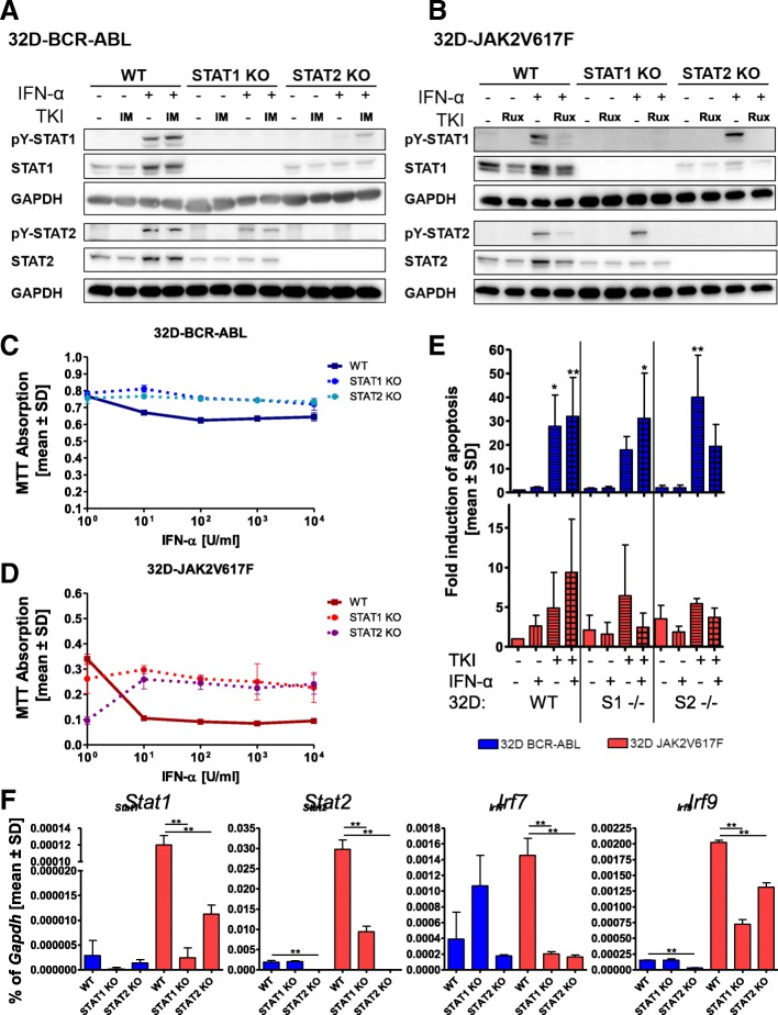
Fig. 3.
STAT1 or STAT2 knockout alters IFNa responsiveness only in JAK2V617F-positive cells. a 32D-BCR-ABL-WT, 32D-BCR-ABL-STAT1ko, and 32D-BCR-ABL-STAT2ko or b 32D-JAK2V617F-WT, 32D-JAK2V617F-STAT1ko, and 32D-JAK2V617F-STAT2ko cells were treated for 4 h with TKI (1 μM imatinib and ruxolitinib, respectively) or IFNa (100 U/ml) or a combination of both. SDS-Page and Western blotting were performed, and the indicated immunostainings were carried out. GAPDH served as the loading control. c 32D-BCR-ABL-WT, 32D-BCR-ABL-STAT1ko, and 32D-BCR-ABL-STAT2ko or d 32D-JAK2V617F-WT, 32D-JAK2V617F-STAT1ko, and 32D-JAK2V617F-STAT2ko cells were treated with increasing concentrations of IFNa, and cell viability was measured by MTT assay. e Indicated 32D cells were treated with TKI (0.5 μM imatinib and 0.1 μM ruxolitinib, respectively) or IFNa (100 U/ml) or a combination of both for 48 h, due to the rapid growth of untreated BCR-ABL-positive cells. PI staining was performed to discriminate between living and dead cells. Mean values ± SD are depicted. f Measurement of Stat1, Stat2, Irf7, and Irf9 mRNA expression in the indicated cell lines. Expression was calculated as a percentage of Gapdh, and the mean values ± SD are depicted. *p < 0.05, **p < 0.01, ***p < 0.001. For all experiments, the respective 32D cells were WEHI starved for 24 h before starting the experiments

