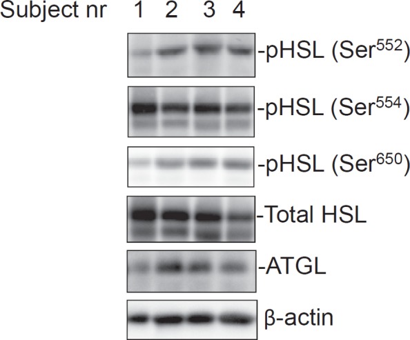
Fig. 1.

Phosphorylated HSL and total HSL and ATGL protein expression levels in white adipose tissue of obese women. Equal amounts of total protein were loaded and separated by SDS-PAGE. Individual p-HSL and total HSL, ATGL and β-actin levels were detected by Western blot and quantified by densitometry as described in material and methods. These representative blots depict the bands detected in four different subjects.
