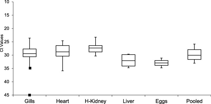
Fig. 2.
Box plot showing the Ct value distribution of each tissue sample tested in the PRV RT-qPCR assay. Gills: n = 21, Heart: n = 21, H-Kidney (Head Kidney): n = 17, Liver: n = 4, Eggs: n = 2, Pooled (tissue pool of gill, heart, spleen, liver, and head kidney): n = 5. The middle lines in each box show the median, and the boxes reflect the quartiles. The error bars indicate the maximum and minimum values within ±1.5 interquartile range of the Ct value distribution. The square (■) represents the minimum and maximum (gills) outliers; the maximum outlier with “No Ct”

