Fig. 4.
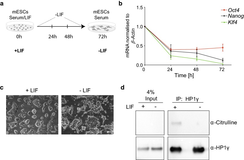
HP1γ citrullination is reduced in differentiating mESCs. a Schematic illustration of differentiation experiments. mESCs were cultured in the presence (+ LIF) or absence of LIF (− LIF) for 24, 48 and 72 h. Experiments were performed at 72 h. b mRNA levels of pluripotency markers in mESCs decrease after withdrawal of LIF. The mRNA levels of the indicated genes were measured by RT-PCR over a course of 3 days after withdrawal of LIF. RT-PCR data were normalised to β-ACTIN mRNA expression, and expression fold change was determined relative to d0 time point using the ddCT method. Bars represent ± SEM, n = 2. c Representative light field microscope images of mESCs before and after 72 h LIF withdrawal, captured with a Leica EC3 camera at × 20 magnification. Scale bars: 100 μm. d Immunoprecipitation (IP) of endogenous HP1γ from nuclear lysates of mESCs cultured +/− LIF for 72 h. IPs were performed with anti-HP1γ antibodies (α-HP1γ) and anti-HA control antibodies and analysed by immunoblotting using an anti-peptidyl-citrulline antibody (α-Citrulline). The same immunoblot was re-probed with an α-HP1γ antibody. 4% input of each IP is indicated. An uncropped image together with two more replicates is shown in Additional file 5: Figure S4A

