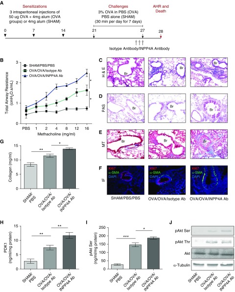
Figure 3.
Treatment with anti-INPP4A antibody aggravates airway inflammation and remodeling in a murine model of AAI. (A) Experimental protocol of antibody-mediated neutralization of INPP4A in a murine model of AAI, developed using sensitization with intraperitoneal injections of 50 μg OVA followed by challenges with 3% aerosolized OVA. (B) Total airway resistance upon methacholine challenge in mice without AAI (sham) and allergic mice (OVA) treated with isotype antibody or anti-INPP4A antibody. (C) Airway inflammation upon antibody treatment as seen by H&E staining. (D) Goblet cell metaplasia as seen by PAS staining. (E) Subepithelial fibrosis as seen by MT staining in lung sections. (F) Representative confocal fluorescence images showing α-SMA (green) and proliferation marker Ki67 (red) in the lung sections of mice treated with isotype or anti-INPP4A antibody (panel elaborated, along with isotype-matched control IgG, in Figure E2E). Nuclei were detected with DAPI (blue). (G) Content of soluble collagen in the lung samples as measured by Sircol assay. (H and I) Expression levels of phosphoinositide-dependent kinase-1 (PDK1) (H) and pAkt Ser in the total lung protein using ELISA (I). (J) Immunoblotting in the lung samples for the expression of pAkt Ser and pAkt Thr with respect to total Akt and α-tubulin. Results are expressed as the mean ± SEM (n = 3–6 mice/group). *P < 0.05, **P < 0.01, and ***P < 0.001 (ANOVA followed by Tukey’s post hoc analysis).

