Figure 6.
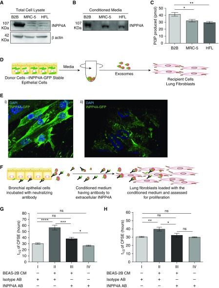
Extracellular INPP4A mediates epithelial–fibroblast communication in the lung and controls proliferation in the recipient cells. (A) Representative immunoblot of INPP4A in lysates of BEAS-2B cells (represented as B2B) and lung fibroblasts (MRC-5 and human lung fibroblast [HFL] cells). (B) Representative immunoblot of INPP4A in the conditioned media of the cells. Normalization by equal seeding density as elaborated in Methods. (C) INPP4 activity in the cell-conditioned media; n = 3–7 per group. (D) Schematic diagram showing isolation of exosomes from MCF-ST cells by differential ultracentrifugation and loading onto MRC-5. (Ei) Representative fluorescent image of MRC-5 cells loaded with exosomes isolated from MCF-ST and imaged after 3 hours. Nuclei are stained by DAPI (blue). (Eii) A three-dimensionally rotated and orthogonally sliced representation of the fluorescence image shows prominent membrane localization of INPP4A-GFP in the recipient MRC-5 cells. (F) Schematic diagram illustrating the incubation of BEAS-2B with antibody for neutralizing secretory INPP4A and loading of this conditioned medium (CM) on lung fibroblasts, followed by checking recipient cell proliferation. (G and H) Half-life of fluorescence intensity of carboxyfluorescein diacetate succinimidyl ester (CFSE) (t1/2) in labeled lung fibroblasts (MRC-5 cells [G] and primary normal human lung fibroblasts [H]) upon treatment of the fibroblast medium directly with isotype or anti-INPP4A antibody, or when fibroblasts were cultured in conditioned media derived from bronchial epithelial cells, with or without neutralization of extracellular INPP4A (n = 5 [G and H]). The half-life of CFSE was measured by monitoring the decrease in CFSE intensity with respect to the harvest time using flow cytometry. Anti-INPP4A antibody used to neutralize extracellular INPP4A; isotype antibody served as the control. Results are expressed as the mean ± SEM. *P < 0.05, **P < 0.01, ***P < 0.001, and ****P < 0.0001; ns = not significant (ANOVA followed by Tukey’s post hoc analysis).

