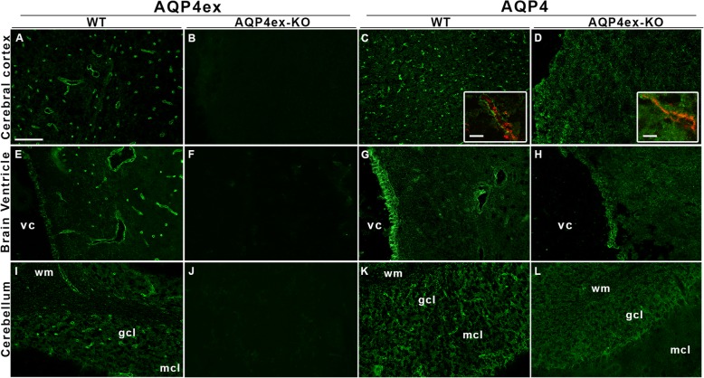
Fig. 3.
Immunolocalization of AQP4 isoforms in WT and AQP4ex-KO mouse brain. The cerebral cortex and the cerebebellum (granular, gcl and molecular cell layer, mcl) are shown together with the inner region (choroid plexus) containing the 4th ventricle cavity (vc) stained with AQP4ex and AQP4 global antibodies (a, e, i). Perivascular staining of AQP4ex is shown in WT mouse (a, e and i), while the signal is absent in KO tissues (b, f, j). The inserts (panel c and d) show a magnified view, obtained by high resolution confocal microscopy, of the perivascular area in WT and KO cerebrum with vessels stained by CD31 antibody (red staining). Note the increased staining of AQP4 on the astrocyte membrane facing the neuropil in the AQP4ex-KO cerebrum. Scale bar 5 μm. A faint staining of AQP4ex was also observed in ependymal cells lining the ventricular cavity (e) and the dense glial processes of the gcl (i) in WT mouse. In AQP4ex mice the staining of AQP4 appeared still present although reduced (h,l). WM, white matter. Scale bar 50 μm

