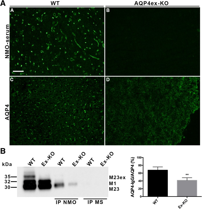
Fig. 7.
NMO-IgG binding in the brain of AQP4ex-KO mouse. a, Immunofluorescence analysis of cerebral cortex of WT and AQP4ex-KO mice stained with NMO-IgG (A,B) and AQP4 (C,D). Note the selective labeling of NMO-IgG to the pericapillary astrocyte processes in WT cortex (A), which is lost in the absence of AQP4ex (B), representative of 14 different NMO-AQP4 positive sera. b, Immunoprecipitation of AQP4 isoforms with NMO sera from WT and AQP4ex-KO cerebrum membrane vesicle extracts. Left, immunoblot probed with AQP4 antibodies showing AQP4 isoforms detected in protein vesicle extracts (WT and AQP4ex-KO) in the first two lanes and in the subsequent lanes the IP with NMO serum and MS serum. Right, densitometric analysis of immunoblot results with 3 different NMO sera. Results are shown as the percentage of AQP4 immunoprecipitated by NMO sera in WT and AQP4ex-KO samples to the total AQP4 immunoprecipitated with anti-AQP4 antibody (AQP4). Mean +/− S.E.M., **p < 0.001)

