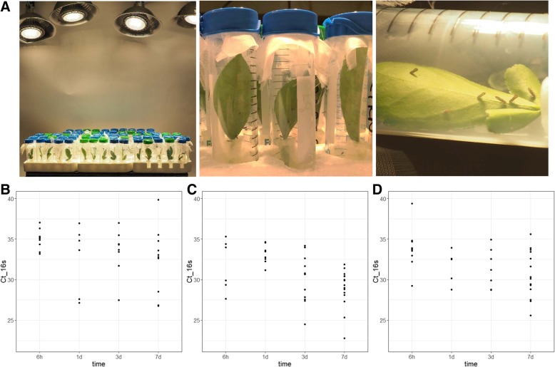
Fig. 2.
Photographs of detached leaf inoculation using Ca. L. asiaticus-infected ACP (a) and the distribution of bacterial titer measured from leaf samples collected at 6 h, 1d, 3d and 7d post ACP exposure (b-d). The experiments were conducted using leaves from citron (b), Duncan (c) and Cleopatra (d). The bacterium was quantified by qPCR amplifying the 16 s rDNA with Las-long primer from 100 ng of citrus DNA template. The cycle threshold values (Ct) were used to generate the dot plots for each time point

