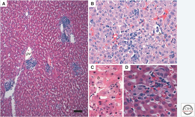
Figure 4.
Acute hepatitis A in a New World owl monkey (Aotus trivirgatus). Staining with hematoxylin and eosin (H&E). (A) Portal tracts are infiltrated with lymphocytes and macrophages primarily. There is no piecemeal necrosis and no derangement of the lobular architecture. Scale bar, 100 µm. bd, bile duct; pv, portal vein; cv, central vein. (B) Inflammation within the parenchyma is characterized by scattered lymphocyte aggregates (a) and accompanied by modest lobular disarray. Ballooning degeneration (b) of hepatocytes is also evident. (C) Ballooning degeneration (b) of hepatocytes generally occurs early in hepatitis A virus infection in owl monkeys. (D) Kupffer cell aggregates (c) may form in the later stages of hepatitis A infection, with or without iron, as seen here.

