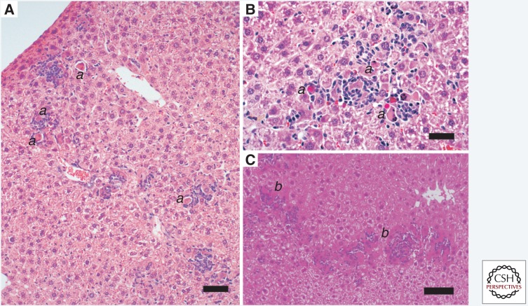
Figure 5.
Hepatitis A in genetically deficient Ifnar1−/− mice that lack expression of the type I interferon (IFN)a/b receptor. Mice were inoculated intravenously with mouse-passaged HM175 virus (Hirai-Yuki et al. 2016). (A) Hematoxylin and eosin (H&E)-stained liver 28 days after inoculation of a 9-week-old female mouse. Serum alanine aminotransferase (ALT) was 57 IU/L, down from 474 IU/L on day 7 postinoculation (upper limits of normal = 30.2 IU/L). Scale bar, 50 µm. Inflammatory infiltrates comprised of foci of lymphocytes and macrophages are scattered throughout the parenchyma, in most cases surrounding remnants of apoptotic hepatocytes (a). Portal inflammation is not evident. (B) Liver collected 41 days after intravenous inoculation of a female Ifnar1−/−Ifngr1−/− double-knockout mouse with third mouse-passage virus; ALT was 201 IU/L. An inflammatory focus is associated with numerous apoptotic hepatocytes (a). Scale bar, 20 µm. (C) Liver from a male Ifnar1−/− mouse collected at necropsy 161 days after inoculation with fifth mouse-passage virus at 9 weeks of age; ALT was 100 IU/L after peaking at 414 IU/L on day 7. Extensive parenchymal infiltrates surround numerous apoptotic hepatocytes (b). Scale bar, 100 µm.

