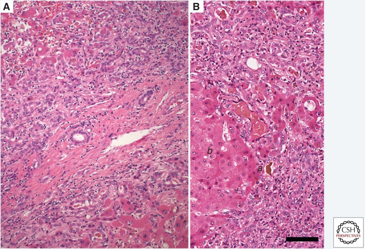
Figure 6.
Hepatitis E in a hepatitis E virus (HEV)-infected patient. (A) Hematoxylin and eosin (H&E) stain. The periportal parenchyma contains a marked ductular reaction characterized by a proliferation of small-caliber ducts lined with pale basophilic epithelial cells extending from the margin of the portal tract. Prominent extracellular matrix separates the ductules. Abutting hepatocytes are in disarray and prominent bile plugs are evident in distended canaliculi and ductules. A mild lymphocytic infiltrate is dispersed through the portal tract and adjacent parenchyma. (B) H&E stain. Higher-magnification image from the same patient. Prominent cholestasis is evident in distended canaliculi with formation of small bile lake at the interface of a prominent ductular reaction on the right and a focus of hepatocytes (a) that are lightly vacuolated and lack normal sinusoidal arrangement. Scale bar, 100 µm.

