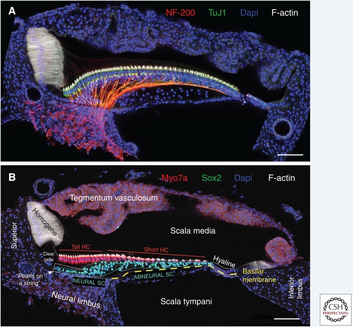
Figure 1.
Cross-sectional anatomy of the chicken basilar papilla (BP). Shown are transverse vibratome sections taken from the middle tonotopic region of the chicken BP, roughly equidistant from proximal and distal ends. Embryonic day 20 (A) and posthatch day 8 (B) were immunostained for (A) neurofilament-200 (NF-200, Sigma) and tubulin β III (TuJ1, EMD Millipore) or (B) myosin-VIIa (Myo7a, Proteus) and Sox2 (Santa Cruz Biotech). Phalloidin labels filamentous (F)-actin and Dapi stains nuclei. Sections were imaged with a Zeiss LSM880 confocal microscope at 40× magnification using Zen Black acquisition software. SC, Supporting cell; HC, hair cell. Scale bars, 50 µm.

