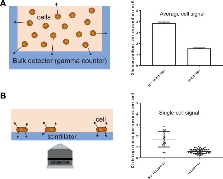
Figure 1.
Bulk and single-cell measurements of FDG uptake. A, Bulk radionuclide counting of cells using a γ counter (schematic) showing the detection of γ rays (arrows) from a suspension of cells inside the γ counter. The FDG uptake in MDA-MB-231 cells is ≈2 times lower in cells treated with αCHC, a lactate export inhibitor. B, Radionuclide counting of single cells using RLM (schematic). Here, the arrows represent β particles emitted following radioactive decay of FDG. As in the bulk experiment, mean FDG uptake is 2 times lower in cells pretreated with αCHC; in addition, quantification of single-cell FDG uptake shows lower heterogeneity when cells are treated with the inhibitor. αCHC, α-cyano-4-hydroxycinnamic acid; FDG, 18F-fluorodeoxyglucose; RLM, radioluminescence microscopy.

