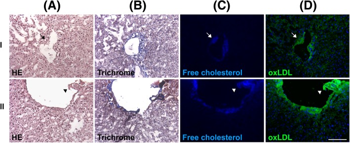
Fig. 2.
Portal venous atherosclerosis in human NAFLD. Two surgical specimens (I and II) were stained with hematoxylin and eosin (H&E) (a), trichrome (b), filipin (c), or an antibody against oxLDL (d). The portal venous plaque showed oxLDL and free cholesterol accumulation. The plaque caused the narrowing of the portal venous lumen (arrows, I) or was protruding and thus ruptured the vein (arrowheads, II). Scale bar: 100 μm

