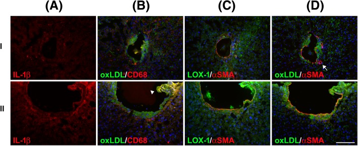
Fig. 3.
Portal venous plaque inflammation marked by macrophage infiltration in NAFLD. The surgical specimens (I and II) were stained with antibodies against IL-1β (a), oxLDL and CD68 (b), LOX-1 and α-SMA (c), and oxLDL and α-SMA (d). Macrophage infiltration (marked by CD68) in the portal vein walls with oxLDL accumulation is shown. A ruptured portal venous plaque showed a particularly high intensity of CD68, LOX-1, and IL-1β signals, suggesting portal venous inflammation (lI, arrowhead). Portal venous atherosclerosis was observed in the layer below vascular smooth muscle (marked by α-SMA) and protruded into the portal venous lumen. The hepatic artery (arrow) was not involved in the oxLDL-related event. Scale bar: 100 μm

