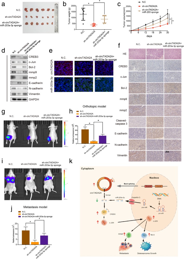
Fig. 8.
CircTADA2A functions as a miR-203a-3p sponge to promote tumorigenesis in vivo. a Nude mice were respectively injected with an equal amount of 5 × 10⁶ stable control cells or cells transfected with circTADA2A shRNA or cotransfected with circTADA2A shRNA and miR-203a-3p sponge subcutaneously. After 30 days, tumors were dissected and photographed. b Tumor weight was calculated on the day mice were euthanized. Data represents the mean ± SEM (n = 6 each group). c Tumor volumes (ab2/2) were recorded every six days since the day when mice were injected the stable OS cells. Data represents the mean ± SEM (n = 6 each group). d Western blotting demonstrated the protein levels of CREB3, c-Jun, Bcl-2, mmp9, mmp2, E-cadherin, N-cadherin and Vimentin in tumors from different groups. e FISH demonstrated the relative expression levels and localization of circTADA2A and miR-203a-3p in the tumors from the mice. Scale bars, 100 μm. f H&E staining and immunohistochemistry (IHC) revealed the structure of OS in mice and relative protein levels of CREB3, c-Jun, Bcl-2, mmp9, mmp2, Cleaved-caspase3, E-cadherin, N-cadherin and Vimentin in tumors of different groups. Scale bars, 100 μm. g & h 143B cells were labeled with both GFP and luciferase. In vivo bioluminescence imaging system showed the orthotopic xenograft tumor in 3 groups of mice injected with 3 different stable 143B cells. Representative images and a histogram are shown (n = 9 each group). i & j Lung metastasis of mice injected with different stable 143B cells via the tail vein was detected using an in vivo bioluminescence imaging system. Representative images and a histogram are shown (n = 9 each group). k Schematic illustration of the circTADA2A/miR-203a-3p/CREB3 axis. Data are from three independent experiments (mean ± SEM) (h and j) or are representative of three independent experiments with similar results (d-f, g and i) (*P < 0.01 vs control or as indicated by Student’s t-test)

