Figure 3.
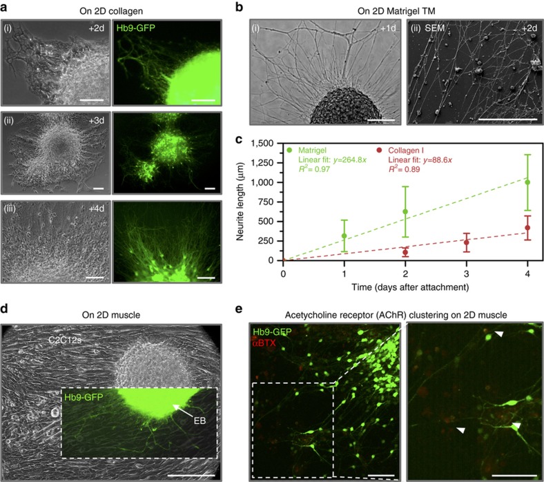
Differentiated embryoid bodies attached readily to 2D ECM-coated substrates such as (a) collagen I (shown after (i) 2, (ii) 3, and (iii) 4 days of attachment) and (b) Matrigel (shown after (i) 1 and (ii) 2 days of attachment). All scale bars represent 100 μm. (c) The EBs extended GFP+ neurites from motor neurons across the surface of the gels upon attachment. The plot represents the mean±standard deviation (n=103–298 neurites from three to seven images per time point). (d) EBs also attached to 2D cultures of differentiating C2C12s and extended neurites across the surface of the myotubes (shown after 1 day of attachment). The scale bar represents 200 μm. (e) Clusters of post-synaptic acetylcholine receptors (AChRs, red, stained with α-bungarotoxin) were visible near the termination of neurite extensions on myotubes 5 days after co-culture. The scale bars represent 100 μm.

