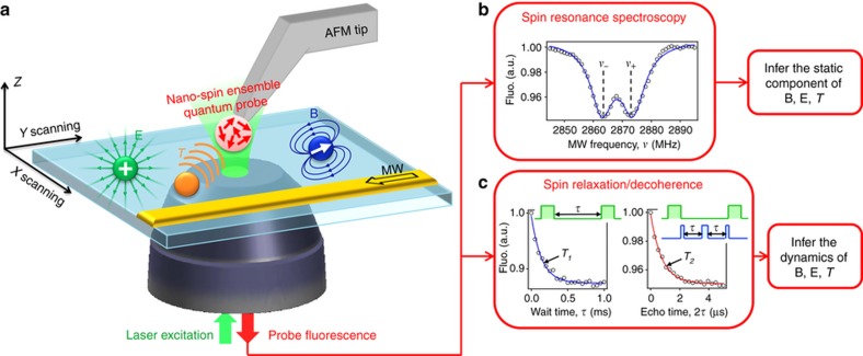
Figure 10.
Schematic of the scanning nano-spin ensemble microscope. (a) Probe consists of a nano-diamond containing a small ensemble of electronic spins (symbolized by the red arrows) grafted onto the tip of an atomic force microscope (AFM). Optical excitation and readout, combined with microwave (MW) excitation, enable quantum measurements of the spin ensemble properties, such as spin resonance, relaxation and decoherence. Scanning the sample relative to the probe produces images of the sample with magnetic (B field), electric (E field) or temperature (T) contrasts, depending on the probing technique used. (b) Optically detected spin resonance spectrum of a nano-diamond on a tip. The solid line is the fit to a sum of two Lorentzian functions centered at frequencies ν±=D±E, providing the zero-field splitting parameters D=2868.3±0.1 MHz and E=5.4±0.1 MHz. (c) Spin relaxation (left) and spin decoherence (right) curve of a nano-diamond on the AFM tip. The inset depicts the sequence of laser (green) and MW (blue) pulses employed. Solid lines are fits to a single exponential decay, revealing a spin relaxation time of T1=142±9 μs and a spin coherence time of T2=780±30 ns. Images reprinted with permission from: Ref. 88, Copyright 2016 American Chemical Society.

