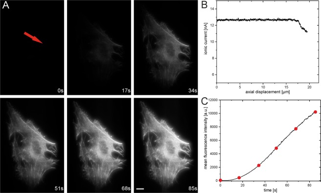
Figure 3.
Nanoinjection of ATTO655-Phalloidin into the cytoplasm of a single living adult human inferior turbinate stem cell (ITSC) after neuronal differentiation (the injection point is indicated by the red arrow). (A) Fluorescence view of the injection of ATTO655-Phalloidin molecules into the cell with an injection voltage of 200 mV applied for 85 s. The images show the delivery of the functionalized fluorescent molecules into the cell by increased fluorescence. The fluorescence is confined within the cell and an instant binding to the actin structure is visible. After ~50 s, the actin structure of the living cell is clearly visible under the applied imaging conditions. The injection was carried out with a concentration of 10−5 M ATTO655-Phalloidin solved in PBS inside the nanopipette. (B) Approach curve of the nanopipette towards the cell. The tip of the nanopipette was placed manually ~20 µm above the coverslip surface. After 17 µm of approach, a first decrease of the ionic current is visible, indicating the contact of the tip with the cell. The approach was stopped manually at 19 µm to avoid breaking of the nanopipette. Afterwards the injection of fluorescent probes began. (C) Average fluorescence intensity of single images taken over the period of injection, indicating the delivery of molecules into the cell. The red dots indicate the time points of the respective fluorescence views in a. Scale bar: 5 µm, images were taken at low wide-field illumination conditions with an integration time of 120 ms.

