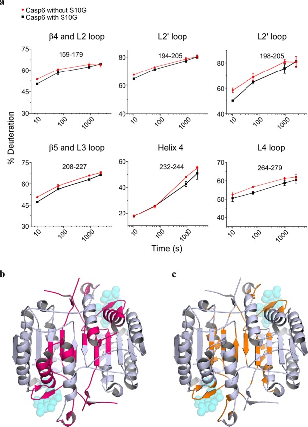
Figure 5.
S10G and Casp6 interaction sites. (a) Representative deuterium uptake plots for peptic peptides of Casp6 incubated without S10G (red) and with S10G (black). Data points represent mean ± SD from three independent experiments. (b) Representative peptic peptides from panel (a) are mapped (pink area) on the CASP6 crystal structure (PDB: 3OD5) and compared to potential binding sites of S10G, as predicted by molecular docking (c, orange area). For indication purposes, Ac-VEID-AFC substrate, as found in CASP6-bound structure, is shown as translucent spheres. Same point of view as in Fig. 3.

