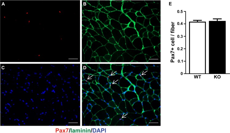
Figure 8.
Satellite cells content after 22 months of Dicer inactivation. Pax7 (Pax7+) immunohistochemistry was used to detect satellite cells in plantaris muscle cross-section. (A) Representative image of Pax 7+ (red loci) co-stained with laminin (green boundary) and DAPI from vehicle-treated (wild-type, WT) plantaris muscle. Scale bar = 50 μm. (B) Quantification of satellite cell abundance in plantaris muscle from WT and tamoxifen-treated (knockout, KO) mice. Data are presented as the mean ± SEM (n = 8–9).

