Figure 1.
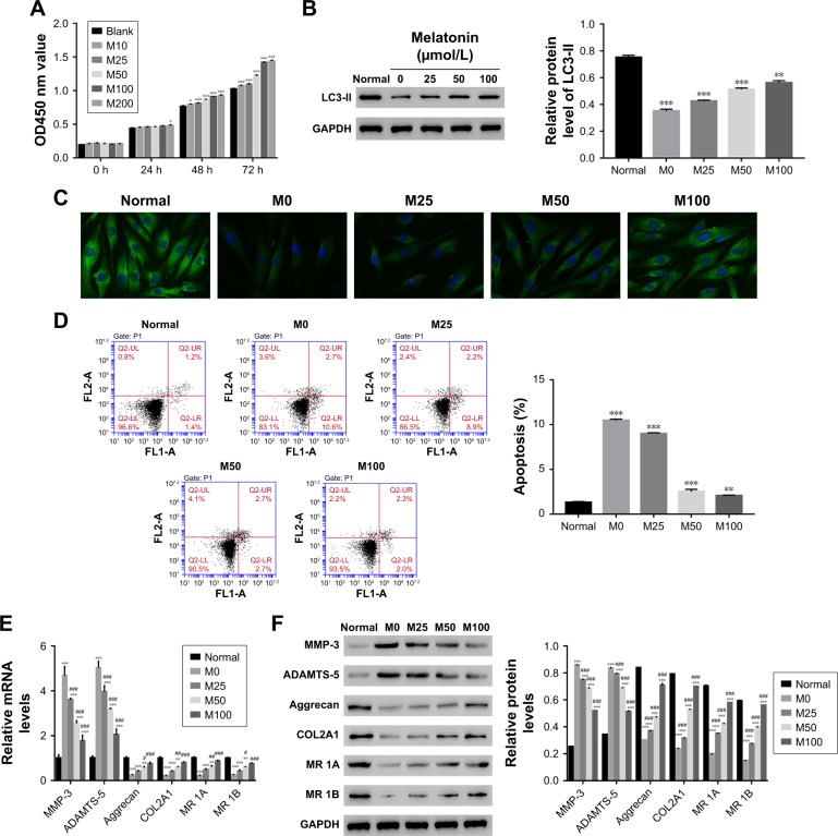
Overload of melatonin stimulated proliferation, induced autophagy, and apoptosis in AF cells of patients with DD. (A) Cell proliferation was detected 0, 24, 48, and 72 hours after treatment in AF cells of patients with DD treated with different melatonin concentrations. (B) Relative protein level of LC3-II in AF cells of patients with DD treated with 0, 25, 50, and 100 µmol/L of melatonin. (C) Representative images of immunofluorescent detection of LC3-II (green) in normal AF cells and AF cells of patients with DD. (D) The cell apoptosis profile of AF cells. (E and F) stand for the mRNA and protein level of MMP-3, ADAMTS-5, Aggrecan, COL2A1, and melatonin receptor 1A/1B in melatonin cultured cells.
Notes: *P<0.05 vs control, **P<0.01 vs control, ***P<0.001 vs control; #P<0.05 vs M0, ##P<0.01 vs M0, ###P<0.001 vs M0. Magnification of the image in (C) is 400×.
Abbreviations: AF, annulus fibrosus; DD, disc degeneration; LC3, light chain 3; M0, M25, M50, and M100, Melatonin of 0, 25, 50, and 100 µmol/L; MR 1A/1B, melatonin receptor 1A/1B.

