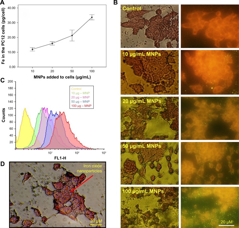
Figure 4.
Internalization of superparamagnetic iron oxide nanoparticles into the PC12 cells. (A) Uptake of iron oxide nanoparticles by PC12 cells 24 hours after exposure to the iron oxide nanoparticles. Iron oxide nanoparticles were added to the media in a dose-dependent manner ranging from 10 to 100 µg/mL. (B) Fluorescent images of incubated cells with the different concentrations of fluorescent iron oxide nanoparticles. (C) Different histograms using Flowing Software (version 2.5.1) were combined: the red curve represents the maximum fluorescence. In this case, PC12 cells were incubated with 100 µg/mL of iron oxide nanoparticles. (D) Prussian blue method: PC12 cells treated with iron oxide nanoparticles were also stained for tracing the iron in the cell cytoplasm. Arrow represents the position of the iron oxide nanoparticles in PC12 cells (dark blue dots).
Abbreviation: MNPs, magnetic nanoparticles.

