Figure 6.
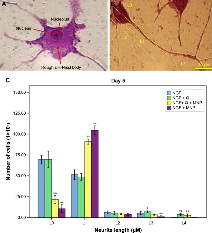
Neurite outgrowth of PC12 cells. (A) Cresyl violet staining: the Nissl substance (rough endoplasmic reticulum) appeared dark blue due to the staining of ribosomal RNA. Magnification ×400. (B) Distribution of the neurite length of PC12 cells 5 days after the induction of differentiation (L0 refers to the cells without neurites; L1 refers to the cells with neurites whose length is shorter than the size of the cell body; L2 refers to the cells with neurites whose length is between the original and twice the size of the cell body; L3 refers to the cells with neurites whose length is longer than twice the size of the cell body). (C) Neurite length of PC12 cells under different treatments. Cells were scored positive if one or more neurites with length >1 cell body diameter was observed. The results presented are the mean ± SD of 10 independent experiments. *P<0.05, **P<0.01.
Abbreviations: NGF, nerve growth factor; Q, quercetin; MNP, magnetic nanoparticle.

