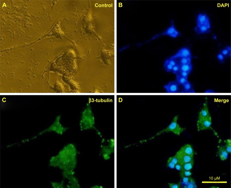
Figure 9.
Immunofluorescence images of differentiated PC12 cells 5 days after treatment with iron oxide nanoparticles. (A) Control, (B) cells stained with DAPI, (C) cells stained with β3-tubulin, and (D) the merge of (B) and (C). Green and blue fluorescence represent β3-tubulin and nucleus, respectively. Nuclei marked with DAPI.

